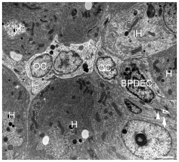
Fig. 7.
Transmission electron microscopy of the liver of a DMN-exposed medaka with oval cell hyperplasia. A row of oval cells (OC) is located between hepatocytes (H) and in contact with a bile preductular epithelial cell (BPDEC) and a cell showing features of hepatocyte differentiation (immature hepatocytes [IH]) including increased cell volume, increased nuclear size, greater number of mitochondria, increased glycogen and development of rough endoplasmic reticulum. A biliary canaliculus formed by a BPDEC and hepatocytes is visible (arrow). Junctional complexes are indicated by arrowheads. Bar, 2 μm.

