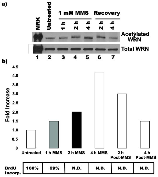
Fig. 4. Kinetics of WRN acetylation.
a) 8-D cells were untreated, incubated with 1 mM MMS for 1, 2 or 4 h, or incubated with MMS for 4 h then released into fresh medium for an additional 2 or 4 h (recovery). For each treatment, cells were harvested and lysates processed for IP with anti-acetylated lysine antibody. The IP products (upper panel) and cell lysates (45 ug each, lower panel) were subjected to Western blotting with anti-WRN antibody. b) Bar graph for WRN acetylation from experiment performed in A, along with table showing, at corresponding time points after MMS treatment, percentages of BrdU incorporation with respect to untreated control (N.D. = not detectable above background)

