
Figure 2.
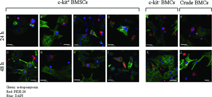
c‐Kit+‐BMSCs express cardiac contractile proteins when co‐cultured with NRVMs: c‐kit+ BMSCs, c‐kit, and crude BMCs were stained with a membrane dye PKH26 before being co‐cultured with NRVMs. The co‐culture was analyzed for the expression of cardiac contractile protein (α‐tropomyosin) at 24 h (A–F) and 48 h (G–L). During the 48 hours of co‐culturing, the c‐kit+ BMSCs expressed α‐tropomyosin. At 24 hours, the majority of the PKH26 stained BMSCs were small round shaped cells and only a few cells had α‐tropomyosin expression (A–D). Some cells were in mitosis, with (C) or without α‐tropomyosin expression (A). At 48 hours, the number of small round cells was decreased (H). The mitotic cells with α‐tropomyosin expression were still present (G) but the majority of the α‐tropomyosin expressing cells were larger (I and J). PKH26 stained c‐kit and crude BMCs were also found to express α‐tropomyosin at 48 hours (K and L); however, at 24 hours, α‐tropomyosin expression were rarely found in these cells (E and F). The percentage of c‐kit+, c‐kit−, and crude BMCs with contractile protein expression at 24 hours was 15.4 ± 1.7, 4.0 ± 1.1, and 6.1 ± 1.2%, respectively. At 48 hours, these percentages increased to 41.1 ± 2.1, 12.3 ± 1.4, and 17.1 ± 1.5%, respectively. Red: PKH26; green: α‐tropomyosin; blue: DAPI.