
Figure 3.
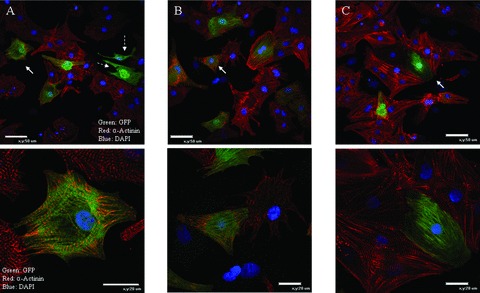
c‐Kit+‐BMSCs differentiate into cardiac myocytes with organized sarcomeres: GFP expressing c‐kit+‐BMSCs were co‐cultured for 48 hours with NRVMs and analyzed for α‐actinin immunofluorescence. 38.6 ± 1.3%(n= 4) of the GFP expressing cells were observed to have organized α‐actinin (A, B, and C). White arrows indicate the cells magnified in the bottom panels. Dotted white arrows indicate GFP expressing BMSC derived non‐myocytes that are not expressing α‐actinin (A). Green: GFP; red: α‐Actinin; blue DAPI.