
Figure 6.
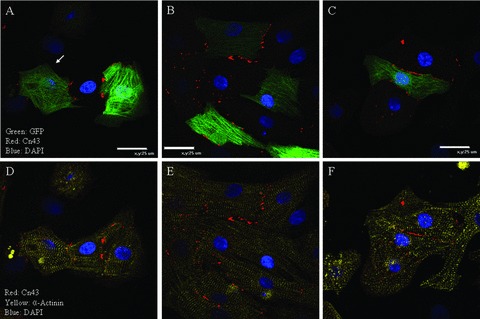
Three representative examples of BMSC‐DMs that form gap junctions with neighboring cardiac myocytes: The GFP expressing BMSCs were co‐cultured with NRVMs. At 48 hours, the co‐culture was analyzed for α‐actinin and connexin 43 (C×43) immunofluorescence. GFP (green) expressing BMSC‐DMs localized C×43 (red) at the cell border with neighboring NRVMs (A, B, and C). Cardiac myocytes were identified by α‐actinin immunofluorescence (yellow: D, E, and F). C×43 was not detected between BMSC‐DMs and non‐myocytes (white arrow: A).