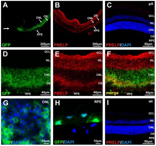
Figure 3. AAV2/8 Mediated Expression of PRELP.
In order to distinguish virus tropism from that of localization of PRELP, AAV2/8-PRELP or AAV2/8-pA was spiked with AAV2/8-GFP. (A) Unidirectional distribution of GFP in the ONL and RPE along the line of the injection (bold arrow) following subretinal injection of AAV2/8-PRELP + AAV2/8-GFP in mice. (B) Cy3 (PRELP) staining in the inner retina and outer segments of photoreceptors. (C) Cy3 labeled section of an AAV2/8-pA injected eye. Nuclei are counterstained with DAPI. (D) Higher magnification of retina from mice following subretinal injection of AAV2/8-PRELP spiked with AAV2/8-GFP. GFP is present in the ONL, outer segments and RPE. (E) Human PRELP is exclusively present in the inner retina and outer segments with a weak signal in the RPE. (F) Merged images of GFP and Cy3 (PRELP) indicating some regions of overlap (yellow) and distinct regions of expression for GFP and PRELP, indicative of PRELP being synthesized in the photoreceptors and RPE but being secreted and localizing to the inner retina and photoreceptor outer segments. (G) Higher power images of GFP/DAPI overlays from the ONL and (H) RPE. (I) Cy3 labeled section of an uninjected wt eye. Nuclei are counterstained with DAPI. RPE; retinal pigment epithelum, OS; photoreceptor outer segments, ONL; outer nuclear layer, INL; inner nuclear layer, GCL; ganglion cell layer.

