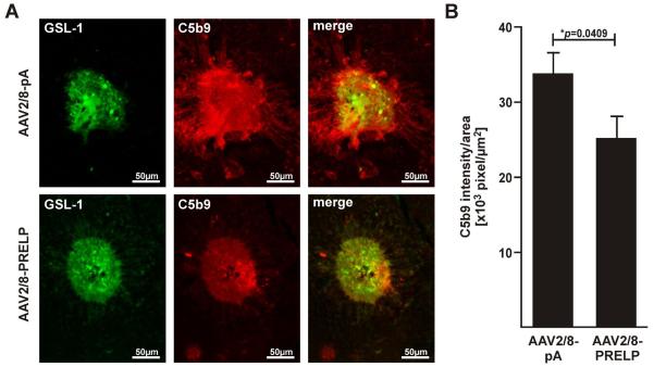
Figure 5. Human PRELP Attenuates Formation of Membrane Attack Complex in Mice.
(A) Representative micrographs of C5b9 labeled laser-induced CNV spots from eyes injected with AAV2/8-pA or AAV2/8-PRELP 7 days post laser. Overlays of GSL-1 and C5b9 stainings of equal sized CNV spots indicating MAC deposition confined within borders of CNV in AAV2/8-PRELP infected eyes, but spread beyond the CNV area in the negative controls. Note that there was variability in GSL-1 staining between laser spots and this was overcome by use of a relatively large sample size. In the above figure, two spots of roughly equivalent GSL-1 staining were selected in order to demonstrate the corresponding differences in C5b9 staining. (B) Graphical plot of mean values±SEM of C5b9 intesity/area ratios in AAV2/8-pA and AAV2/8-Prelp injected eyes. Average ratio was significantly reduced by 25.5±12.3% (*p=0.0409) in AAV2/8-PRELP infected eyes. Studies were performed twice with 5 mice in each group (n=10 mice/ 20 eyes; AAV2/8-pA: 61 laser spots, AAV2/8-Prelp: 55 laser spots).

