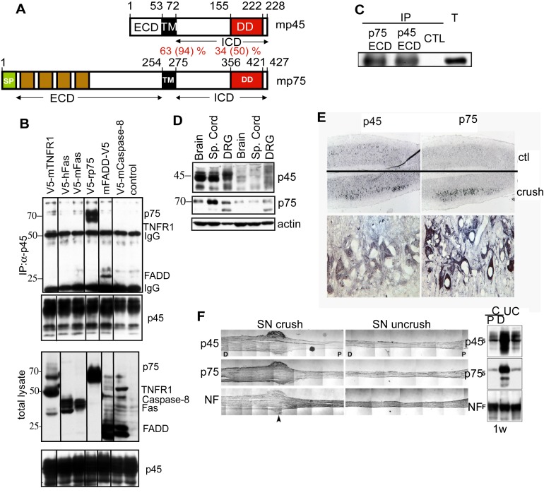
Figure 1. Interactions between p75 and p45.
(A) A schematic diagram showing domains of p45 in comparison with p75. TM, transmembrane domain; DD, putative death domain; PDZ, putative PDZ binding domain. The degrees of identity and homology in amino acid residues between mouse p75 and p45 are shown in percentages. (B) P45 forms a complex with FADD and p75. V5-tagged TNFR1, human Fas, mouse Fas, p75, FADD, or Caspase-8 was transfected into CrmA/Flag-p45/293 stable cells. The lysates were immunoprecipitated with anti-Flag antibodies and immunoblotted with anti-V5 antibodies. (C) P7 cerebellum extracts were immunprecipitated with anti-p75 ECD antibody (9651) or anti-p45 ECD antibody (6750) followed by immnuoblotting with anti-p75 antibody (Buster). T, total lysate. (D) Western blotting analysis of p45 and p75 expression in the brain, spinal cord, and DRG of P0 and adult mice. (E) Increased expression of p45 and p75 in the spinal cord and sciatic nerve following sciatic nerve injury. (Top) Spinal cord sections were immunostained with anti-p45 or -p75 antibodies. p45 and p75 immunoreactivities were markedly increased in the ipsilateral side as compared to the contralateral side. Higher magnification indicated that expression of p45 and p75 is increased in motor neurons. (F) Longitudinal sections of crushed and uncrushed sciatic nerves were immunostained with antibodies against p45, p75, or neurofilament. The levels of p45 and p75 were markedly increased in the distal (D) portion of sciatic nerves as compared to the proximal (P) end and the uncrushed (UC) nerve.

