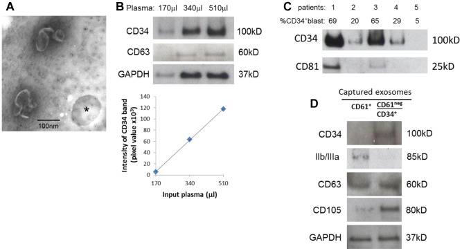
Figure 5. Capture of CD34+ blast-derived exosomes directly from AML patients' plasma.
A) Negatively stained electron microscope images of exosomes captured on CD34 microbeads (* shows a vacant microbead). B) Increasing AML plasma volumes were used for capture of CD34+ exosomes by microbeads and recovered exosomes were studied by Western blots. The graph shows a linear relationship between the input plasma volumes and pixel densities of captured and blotted CD34+ exosomes. C) Exosomes were captured from plasma samples obtained from five CD34+ AML patients and were analyzed by Western blots. The percentage of leukemic blasts in the peripheral circulation of each of the patients is shown. CD81 serves as the exosome marker. D) Removal of platelet-derived exosomes from plasma using anti-CD61 Ab-coated microbeads prior to capture of CD34+ exosomes. Exosomes captured with CD61 microbeads (left: CD61+) and CD34+ exosomes captured after removing CD61+ exosomes (right: CD61neg/CD34+) are shown in a representative Western blot of three evaluated.

