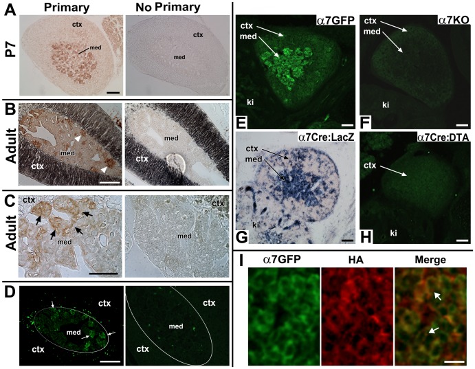
Figure 1. Specificity of α7G detection in the adrenal gland.
A. Transverse sections from a post-natal day 7 (P7) α7G mouse adrenal gland. Anti-GFP (Primary) labeling using the immuno-peroxide method reveals cells localized throughout the adrenal medulla (med) that exhibit expression. Cells in the adrenal cortex (ctx) were not labeled. On the right is a similar section that was processed but the antibody to GFP was omitted (No Primary). B. Transverse sections from an adult α7G adrenal gland processed for GFP immunoreactivity as in A. On the right an adjacent section was processed without addition of anti-GFP primary antibody. Arrow heads identify cells immunopositive for GFP signal which tend to be in clusters near the boundary of the medulla and cortex (darkly colored in this preparation). C. Increased magnification of the adult adrenal gland medullary region showing immune-reactivity towards GFP. The arrows point to cell clusters expressing GFP indicative of α7G expression. The section to the right was again processed the same way but with omission of primary antibody. D. Transverse sections from an adult α7G mouse adrenal gland were processed using immunohistochemistry to reveal anti-GFP immunofluorescence. On the right is a section processed without addition of anti-GFP primary antibody. Arrows identify α7G cells immunopositive for GFP signal. The white oval outline approximates the boundary between the med and ctx. Note the similarity between the immunofluorescence pattern of GFP expression in cell clusters near the medulla-cortex boundary that closely resemble the pattern of immune-staining seen in B and C using the immuno-peroxide method. E–H. Additional tests confirm the specificity for measurement of the α7G expression pattern in the adrenal gland. E. Sections were taken from E14.5 embryos of various genotypes related to α7 expression. First, immunofluorescence to α7G (α7GFP) is revealed using and anti-GFP primary antibody. The anti-GFP signal is prominently expressed in chromaffin cells of the developing adrenal medulla (med), but it is essentially absent from the adrenal cortex (ctx). The kidney is identified (ki). F. A similar section was prepared from an α7 knock-out (α7KO) adult mouse and processed for anti-GFP immunofluorescence. No detectable GFP signal was observed. G. Another independent method to measure α7 expression was tested in an E14.5 embryo from the α7Cre to a Rosa26-loxP(LacZ) reporter mouse cross (α7Cre:LacZ; for details see [28]). This embryo exhibits beta-galactosidase staining consistent with chromaffin cell expression. H. Sections prepared from E14.5 embryo of the α7Cre x Rosa26-LoxP(diphtheria toxin (DTA) cross to conditionally ablate (α7Cre:DTA; see [28]). Anti-GFP immunofluorescence detects no α7G and there is no defined adrenal medulla (compare with E). I. In an E18.5 α7G embryo the co-labeling for GFP (α7GFP; green) and the α7-associated hemagglutinin epitope tag (HA; see [28]) reveals GFP (green) that diffuses throughout the cytoplasm but does not enter the nucleus. The HA immunostaining (red) is located on the cell surface of the GFP expressing cells usually in a punctate pattern (identified by arrows). For A–H the Bar = 100 µm and for I the Bar = 30 µm.

