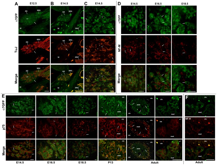
Figure 5. Adrenal gland innervation and expression of α7G.
Adrenal glands at various times of development as indicated were labeled for co-expression of α7G as measured by anti-GFP immunofluorescence (α7GFP, green) and various peripheral nerve markers as indicated (red). A) The same section from Figure 2a for the E12.5 adrenal gland is shown with co-labeling for immature neuronal marker, beta-III tubulin (TuJ). TuJ labeling reveals penetration of efferents into the developing adrenal gland (AG) and staining in the suprarenal sympathetic ganglion (SRG). With rare exceptions (arrows), there is almost no overlap in cells or processes expressing these respective markers. The kidney is identified (ki). B) The E14.5 AG exhibits consolidation of the TuJ staining into fibers that localize to the site of α7G labeled cell accumulation. Rare cells are labeled by both α7G and TuJ (black filled arrow). Note the coincident staining between these markers in the DRGs, but only very rare TuJ identified filaments in the kidney. C) Increased magnification of the E14.5 adrenal gland shown in B. In these images the cell exhibiting both α7G and TuJ labeling is identified by the black arrow. Aggregated α7G labeled cells with the presence of extensive TuJ-labeled afferents is identified for two of these clusters by arrows. Also present are α7G-labeled cells with no apparent association with TuJ labeled afferents (arrow heads). In some instances the TuJ identified afferents form fine fibers with varicosities that appear to wrap α7G stained cells (black filled arrow with asterisk). D) Shown are images from the AG medulla at the developmental stage indicated double labeled for α7G or neurofilament-M (NF-M). Arrows identify cells expressing α7G that appear to also be in association with NF-M stained afferents. Note the consolidation and apparent trimming of NF-M afferents and their association with clusters of α7G cells. E) Sections of the AG at the indicated developmental time are double labeled with α7G and the neurotrophin receptor subunit p75. Although no co-labeling of cells or processes with α7G and p75 was identified, there is extensive association between α7G cells and p75 labeled afferents that persists into the post-natal period (P13 shown) and becomes refined to include almost preferentially cell clusters of the adrenal medulla identified by the α7G-labeled cell clusters in the adult (arrows). This is particularly evident in the adjacent panels showing these clusters at increased magnification. There are however, some cell clusters in the adult that exhibit weak or no α7G immunostaining that are innervated by p75 stained fibers (asterisk). Occasionally p75-labeled processes also extend into the adrenal cortex (open arrow). These processes are not detected by anti-GFP staining. F) The adult immunostaining by the neurofilament protein-H (NF-H) exhibits less specificity for afferents interacting with α7G stained cells when compared to those afferents identified by p75. Abbreviations: AG, adrenal gland; ctx, adrenal cortex; ki, kidney; med, adrenal medulla; SRG, suprarenal ganglion. Scale bar = 100 µm (A,B, E adult) or 50 µm.

