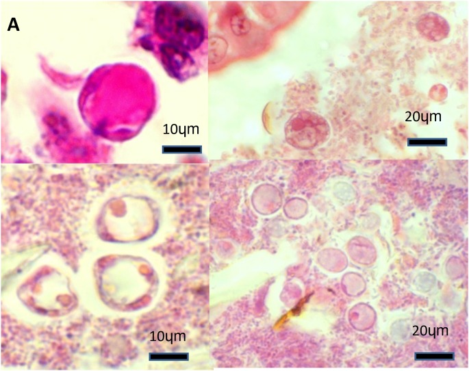
Figure 2. Histological images of Blastocystis organisms in the porcine intestinal biopsies. Haematoxylin and eosin.
(A) Caecum from a commercial pig: a Blastocystis vacuolar form amongst intestinal lumen material (100X). (B) Colon from a research pig: several Blastocystis granular forms found near the tip of the intestinal epithelium (40X). (C) Colon from a commercial pig: three Blastocystis vacuolar forms of amongst luminal material (100X) (D) Colon from a commercial pig: several vacuolar Blastocystis organisms scattered amongst intestinal lumen material (40X).

