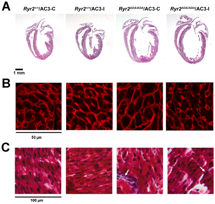
Figure 3. Morphological analysis of wild type and mutant hearts.
(A) Representative sections of hearts from the indicated mice stained with hematoxylin and eosin. (B) Left ventricle sections were stained with TRITC-conjugated wheat germ agglutinin. Cross-sectional areas (µm2, n = 10 for each heart) were (in parentheses) for 2 Ryr2+/+/AC3-C (67.9±2.3), 3 Ryr2+/+/AC3-I (66.8±2.8), 4 Ryr2ADA/ADA/AC3-C (96.0±3.0) and 4 Ryr2ADA/ADA/AC3-I (86.9±3.3) hearts. The significantly increased cross-sectional area in Ryr2ADA/ADA hearts compared to Ryr2+/+ hearts was not significantly altered by AC3-I compared to AC3-C using one way ANOVA. (C) Papillary muscle stained with Masson Trichrome. Arrows indicate collagen deposits.

