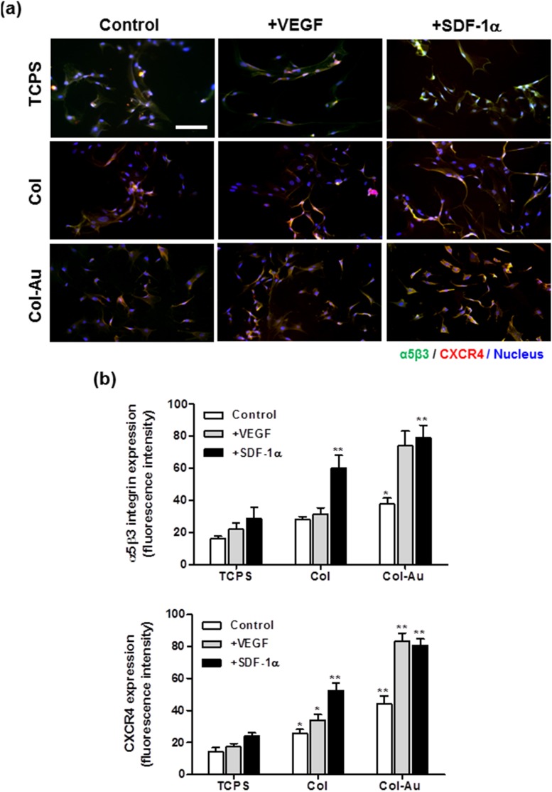
Figure 8. The expression of αVβ3 integrin and CXCR4 for MSCs on different materials at 48 h of incubation and for those treated with either VEGF (50 ng/ml) or SDF-1α (50 ng/ml) in culture media.
(a) MSCs were immunostained by the primary anti-αvβ3 integrin antibody and primary anti-CXCR4 antibody and conjugated with FITC-immunoglobulin secondary antibody (green color fluorescence), Cy5.5-conjugated immunoglobulin secondary antibody (red color fluorescence). Cell nuclei was stained by DAPI. Scale bar = 100 µm. (b) αvβ3 integrin and CXCR4 expressions were quantified based on fluorescence intensity. *p<0.05, **p<0.01: greater than control (TCPS).

