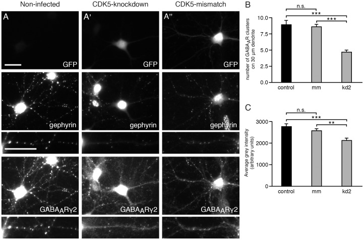
Figure 2. CDK5 knockdown correlates with reduced numbers of GABAA receptor clusters containing γ2-subunits.
Hippocampal neurons (div14) were immunolabeled with anti GFP antibodies to detect infected neurons (upper panel), with phosphospecific anti gephyrin mAb7a antibody (middle panel), and with anti-GABAA receptors γ2-subunit of (lower panel). (A) Non-infected cells; (A') CDK5-kd2 knockdown; (A'') control shRNA (CDK5-mismatch). Neurons were infected with the indicated viruses at div6. The scale bar represents 15 µm. (B) Quantification of the number of GABAA receptor γ2 puncta on three proximal dendritic segments of 30 µm. 20 or 18 cells from n = 3 independent cultures of control or mismatch neurons and 25 cells (69 dendrites) from n = 4 independent cultures of kd2-infected neurons, mean ± SE. ANOVA with post-hoc test, *** P<0.001. (C) Quantification of the relative fluorescence intensities of GABAA receptor γ2 puncta. Mean ± SE. Each value was calculated from data from the same cell numbers as in B from n = 3 independent cultures. ANOVA with post-hoc test: *** P<0.001, ** P<0.01.

