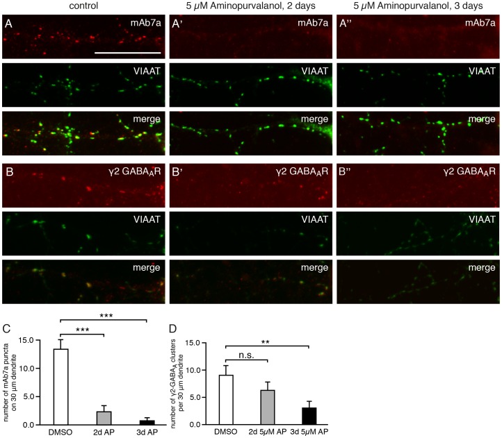
Figure 4. Gephyrin and GABAA receptor γ2 puncta are reduced upon CDK5/2/1 inhibition.
(A) Hippocampal neurons were double-immunolabeled for gephyrin with antibody mAb7a (red, upper panel) and with anti-VIAAT antibody (green, middle panel). Superposition of both immunolabelings (lower panel, merge). Neurons were fixed and immunolabeled at div16. (A) Control cells, non-treated; (A') cultures treated with aminopurvalanol A (5 µM) for 2 days; (A'') cultures treated with aminopurvalanol A (5 µM) for 3 days. Note a higher number of VIAAT-opposed gephyrin puncta (yellow, merge) under A, compared to A' and A''. The scale bar represents 15 µm. (B) Hippocampal neurons were double-immunolabeled for GABAA receptors with anti-γ2-subunit antibody (red, upper panel) and with anti-VIAAT antibodies (green, middle panel). Superposition of both immunolabelings (lower panel, merge). Neurons were fixed and immunolabeled at div16. (A) Control cells, non-treated; (A') cultures treated with aminopurvalanol A (5 µM) for 2 days. (A'') cultures treated with aminopurvalanol A (5 µM) for 3 days. Note a higher number of VIAAT-opposed γ2-subunit puncta (yellow, merge) under B, compared to B' and B''. (C) Quantification of the number of gephyrin (mAb7a) puncta. Quantification was done with about 10 cells from 4 independent cultures. Mean ± S.E.; ANOVA with post-hoc test: * P<0.05. (D) Quantification of the number of GABAA receptors with anti-γ2-subunit antibody. Mean ± SE. Quantification was done with 49 cells from n = 4 independent cultures (control), 34 cells from n = 3 independent cultures (2 days) and 18 cells from n = 3 independent cultures (3 days). ANOVA with post-hoc test: ** P<0.01.

