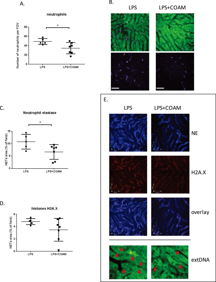
Figure 4. Effects of COAM co-treatment on LPS-induced systemic inflammation in the liver.
(A) Co-application of COAM (2 mg/mouse) with intraperitoneally administrated LPS (1 mg/kg) decreases neutrophil infiltration to the liver at 4 h of inflammation; (B) representative images of neutrophils present in the liver sinusoids of LPS- and LPS plus COAM-treated mice (green cells – autofluorescent hepatocytes; 20x; scale bars represent 50 µm). Quantification of extracellular neutrophil elastase (C) and histone (D) within the livers of LPS and LPS+COAM-treated animals (mean area of staining per 20×FOV ± SD; scale bars represent 45 µm). Intravital visualization of NET deposition in the liver vasculature of LPS-treated and LPS plus COAM-treated mice (E). Staining for extracellular neutrophil elastase (NE) and histone illustrates clear deposition of these characteristic molecules of NETs in the liver after either treatment. In addition, overlay of histone and elastase staining is shown. Staining for extracellular DNA is presented with a higher magnification to clearly picture Sytox green deposition along the liver sinusoids; areas of the extDNA deposition are marked with red arrows. Neutrophil, elastase and histones were measured in five FOV/mouse, n = 5–7 animals per group; *P<0.05.

