Figure 5.

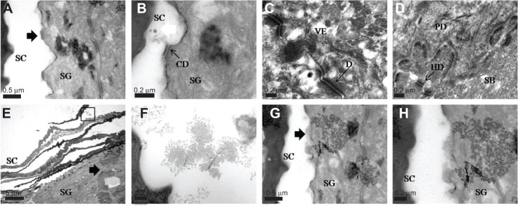
Transmission electron microscopy images of pig skin treated with ultradeformable liposomes, obtained from ultrathick section of pig skin treated with ultradeformable liposomes at 4 hours.
Notes: (A) Corneodesmosome degradation (black arrow) between SC and SG; scale bar represents 0.5 μm. (B) Corneodesmosome degradation (black arrow) and disintegrated particle (white arrow) between SC and SG; scale bar represents 0.2 μm. (C) Desmosome in viable epidermis; scale bar represents 0.2 μm. (D) Hemidesmosome at stratum basale and papillary dermis; scale bar represents 0.2 μm. (E) Ultradeformable liposome penetration into the skin (black arrow); scale bar represents 5 μm. (F) Magnification of marked area in (E); scale bar represents 0.2 μm. (G) Disrupted cell membrane (black arrow) from penetration of intact vesicles into SG; scale bar represents 0.5 μm. (H) Magnification of (G); scale bar represents 0.2 μm.
Abbreviations: SC, stratum corneum; SG, stratum granulosum; SB, stratum basale; PD, papillary dermis; D, desmosome; CD, corneodesmosome; HD, hemidesmosome; VE, viable epidermis.
