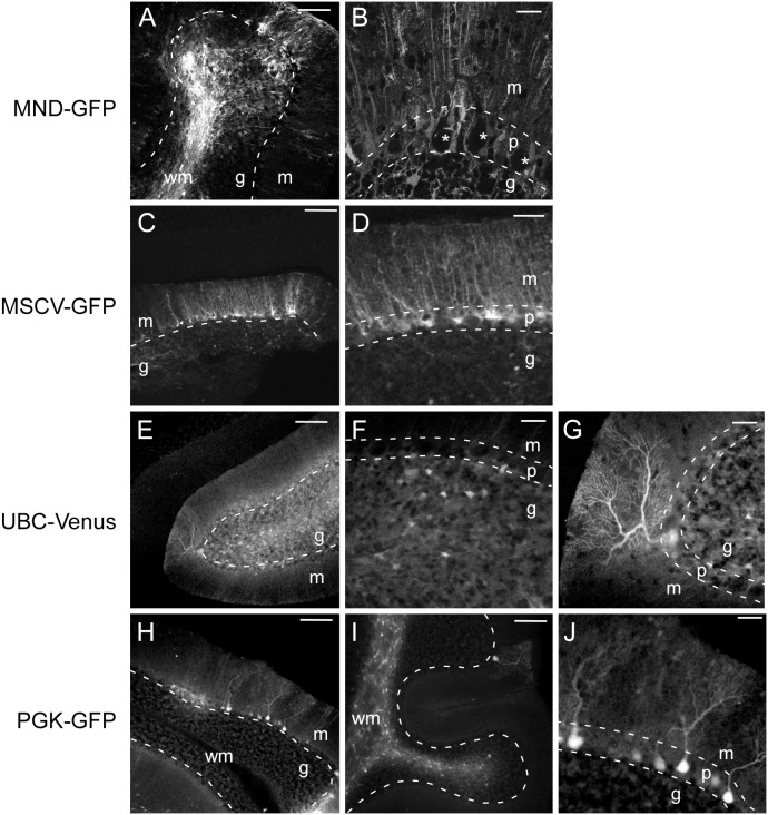
Figure 3. Expression of reporter genes in cerebellar cells transduced with lentiviral vectors under various promoters.
Representative confocal images of sagittal cerebellar sections from mice 7–14 days following intracerebellar injection of lentiviral vectors with indicated promoters. Dotted lines demarcate the border between cerebellar cortex layers. In low magnification images (B, D, F, G, and J), the line is drawn between Purkinje and granule layers. In high magnification images (A, C, E, H, and I), two lines are drawn to separate the Purkinje layer from the molecular layer and granule layer. A. Widespread GFP expression in a cerebellar lobe injected with MND-GFP. B. Single confocal section of MND-GFP transduced cerebellum at higher magnification showing absence of GFP expression in Purkinje neuron somata (asterisks). C. Low magnification of cerebellum injected with MSCV-GFP. D. High magnification of cerebellum injected with MSCV-GFP demonstrating GFP expression in small cell bodies in the Purkinje layer with radial processes extending to the pial surface, characteristic of Bergmann glia. E. Venus expression in a cerebellar lobe injected with UBC-Venus. F, G. High magnification of UBC-Venus infected cerebellum shows venus expression in multiple small cells in the granule layer (F) and a single Purkinje neuron (G). H, I. GFP expression in cerebellar lobes of two animals injected with PGK-GFP. Several GFP-expressing Purkinje neurons are visible in H, whereas most GFP-expressing cells in I are in the white matter, with a single GFP-positive Purkinje neuron. J. High magnification view of GFP-positive Purkinje neurons from H. Abbreviations: m = molecular layer; p = Purkinje layer; g = granule layer; wm = white matter. Scale bars, 25 µm (B, D, F, G, J), 100 µm (A, C, E, H, I).

