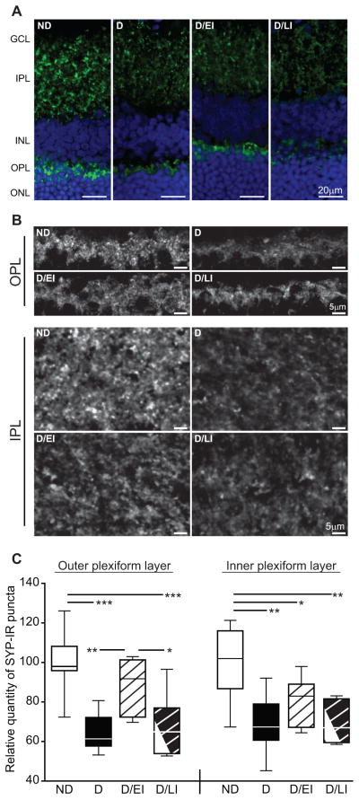
Figure 6. Retinal synapse quantitation.
A) Retinal synaptophysin immunoreactivity (SYP-IR), assessed as a marker of synaptic terminals, was distributed throughout IPL and OPL as punctate staining. There was qualitatively less immunoreactivity in both plexiform layers of uncontrolled diabetic (D) rats compared to non-diabetic (ND) controls. This decreased in qualitative immunoreactivity was attenuated by insulin replacement in the diabetic/early intervention (D/EI) group, while the appearance of the diabetic/late intervention (D/LI) group was closer to that of the D animals. B) High-magnification images of retinal plexiform layers demonstrate a decrease in the intensity of SYP-IR as well as an apparent reduction in the number of SYP-IR puncta in the IPL and OPL of D rats, with similar profiles observed in D/LI group. SYP-IR in the OPL of the D/EIgroup was qualitatively similar to that of ND controls, although reduced staining was apparent in the IPL. C) Automated quantitation of SYP-IR puncta numbers revealed significant decreases after twelve weeks of uncontrolled diabetes compared to age-matched ND controls. Insulin treatment in the D/EI group prevented synapse loss in the OPL, but not the IPL. SYP-IR puncta numbers in the D/LI group were reduced to a similar extent as that observed in D rats, suggesting that six weeks of insulin replacement following six weeks of uncontrolled diabetes is not sufficient to prevent or reverse loss of SYP-IR puncta. ANOVA/SNK, *p<0.05, **p<0.01, ***p<0.001, cohorts 1 and 2 (n=8/group).GCL – ganglion cell layer, IPL – inner plexiform layer, INL – inner nuclear layer, OPL – outer plexiform layer, ONL – outer nuclear layer.

