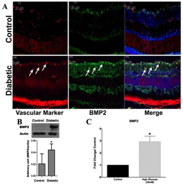
Figure 2. Increased BMP2 expression in Retina of Diabetic mice.
A) Immunofluorescence of BMP2 in normal and diabetic mouse retina. There is marked increase in the expression of BMP2 (Green) in diabetic mouse retina compared to the normal retina (Control). BMP2 is localized mainly in relation to retinal vasculature (Red) (Arrows). B) Western blot analysis of BMP2 in normal and diabetic mice retina showing upregulation of BMP2 in retina of diabetic mice compared to control group (n=5). (C) BMP2 ELISA of conditioned media of high glucose (30mM) treated hRECs for 5 days. Results are plotted as fold change/control and show a significant upregulation of BMP2 as a result of HG (2.9 ±0.4, pvalue: 0.02, n=4) (*:pValue < 0.05 versus control group).

