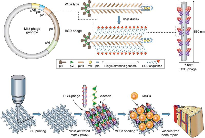
Figure. 1. An overview of VAM in vascularized bone repair.
Top: RGD peptide is fused to the solvent-exposed terminal of each copy of major coat protein (pVIII) constituting the side wall of filamentous phage by inserting gene into gene VIII, generating RGD-phage. Bottom: RGD-phage nanofibers (negatively charged) are integrated into a 3D printed bioceramic scaffold along with chitosan (positively charged), which electrostatically stabilizes the phage nanofibers inside the scaffold. The resultant scaffold is seeded with rat MSCs and then implanted into bone defect. The presence of RGD-phage in the scaffold induced the formation of new bone filled with new blood vessels.

