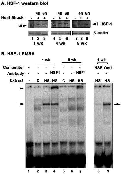
Fig. 6. Activation of HSF-1 in response to heat shock at different ages in D. pulicaria.
A. Western blot analysis. D. pulicaria (lake XVI-clone 11) were subjected to heat shock at 32° C for 30 minutes, allowed to recover for indicated time periods, and protein was extracted. Western Blot analysis was performed using 50 μg of total protein with anti-HSF-1 antibody. The recovery periods after heat shock are as indicated above the lanes and the age of the Daphnia are indicated in weeks below the panels. Blots were re-probed with anti-β actin antibody to ensure even loading. Arrowhead marked “ui” indicates HSF-1 position from non-heat shocked samples, and an arrowhead marked “i” indicates upward mobility shift of HSF-1 in heat shocked samples. B. Electrophoretic mobility shift analysis. EMSA was performed using nuclear extracts prepared from D. pulicaria (lake XVI-clone 11) with (HS) or without heat shock (C) at different ages as indicated. 5 μg of nuclear extracts were incubated with P32-labeled HSE probe. Above each lane, the age of the Daphnia in weeks, addition of a competitor (HSE: specific, Oct1: nonspecific), or addition of an antibody (HSF-1) is as indicated. The ‘-’ lane indicates probe alone lane without any added nuclear extract. Arrows indicate the position of HSF-1 containing complex, and arrowhead indicates position of complex after antibody super-shift. Specific and non-specific non-radioactive competitor oligonucleotides were used in 50-fold molar excess to confirm the specificity of the bound complex.

