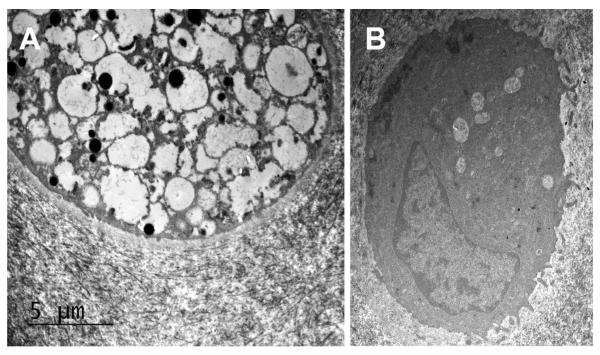
Figure 6.
Transmission electron micrographs of chondrocytes taken from cartilage at baseline (A) and following treatment with IA-rhIDUA (B). Notice the large amount of lysosomal storage vacuoles in the untreated chondrocyte. The treated chondrocyte demonstrates a small number of residual lysosomal storage vacuoles but has normal-appearing cytoplasm and morphology.

