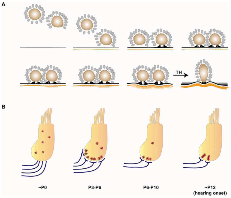
Fig. 2. Development of the mouse inner hair cell (IHC) ribbon synapse.
A. Presynaptic ribbon complexes are formed in IHCs and descend toward the basal cell membrane. Presynaptic densities are anchored by two rodlets to the membrane thickening. Synaptic clefts begin to be filled with a dense filamentous matrix. Subsequently, postsynaptic densities (PSDs) are assembled at sites apposing presynaptic active zones (Sobkowicz et al., 1986). In the mature ribbon synapse, the presynaptic complex is attached to the active zone by a single curved density and the PSD forms a concave shape that exceeds the territory of the presynaptic active zone (Sobkowicz et al., 1982). During early development, ribbons are round and tend to occur in clusters. Mature ribbon synapses are elliptical, and each individual afferent is juxtaposed to a single ribbon. This maturation process depends on thyroid hormone. B. Ribbons are gradually localized to the basolateral surface of the IHC in response to the innervation of SGN neurites during perinatal stages. After the first postnatal week, pruning, retraction and refinement of afferent fibers result in reduction of ribbon synapse number. Concurrently, clusters of ribbons consolidate. After hearing onset, each individual afferent terminal is apposed to a single ribbon.

