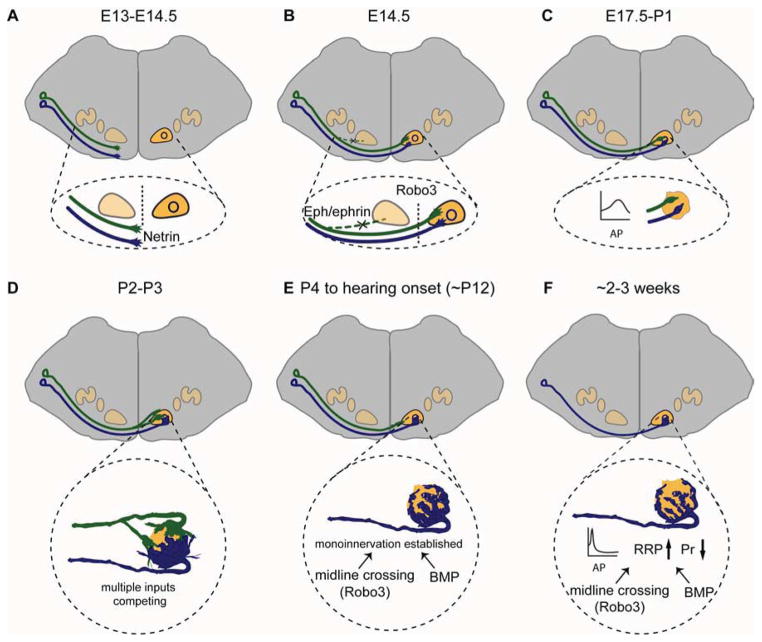
Fig. 4. Major stages in the development of the calyx of Held synapse in mice.
A. Around E13 to E14.5, GBC axons are attracted to the midline by Netrin-1/DCC signaling. B. By 14.5, the majority of GBC axons have crossed the midline of the brainstem due to the expression of Robo3, which prevents premature Slit responsiveness in pre-crossing axons. In parallel, EphB2/Ephrin-B2 signaling suppresses the formation of aberrant ipsilateral VCN-MNTB projections and is required for strictly contralateral VCN-MNTB projections. C. By E17, the earliest synaptic contacts between GBC axons and MNTB principal neurons are established. At P0 and P1, only a few small axosomatic contacts are formed on MNTB neurons. During this time, the presynaptic action potential (AP) has a long duration and small amplitude. D. Around P2-P3, presynaptic endings have formed large cup-shaped swellings called protocalyces, which contain many collaterals and are structurally dynamic. MNTB neurons receive inputs from several calyces at this stage. E. During the first and second postnatal weeks, collaterals are retracted and parts of the calyx become thinner and more intricate. Synaptic competition between multiple calyceal inputs is largely resolved, such that the strongest calyceal input wins, with monoinnervation of nearly all MNTB neurons by this stage. F. Adult calyx of Held synapses show a highly elaborate structure. Presynaptic AP kinetics are faster, with a shorter duration and larger amplitude. In addition, the readily releasable vesicle pool (RRP) size is increased and release probability (Pr) is decreased. Both the morphological and functional maturation of the calyx of Held require BMP signaling and Robo3-mediated axon midline crossing. The timeline is indicated as embryonic and postnatal ages in mice.

