
Figure 1.
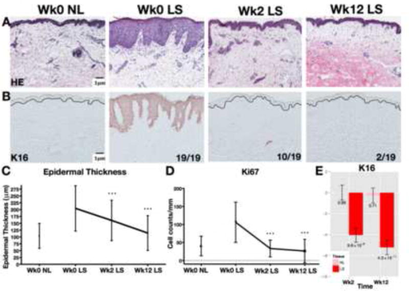
Representative staining with H&E (A) and proliferation marker K16 (B). All 19 LS samples were K16-positive at wk0 and only 2/19 LS samples retained positivity at wk12. Significant decreases in epidermal thickness (C), Ki67+ cell counts (D), and K16 mRNA expression (E) with CsA. Wk/week; LS/lesional; NL/non-lesional; FCH/fold-change; Mean±SD(C–D); Mean±SEM(E); post-versus-pre p-values *p<0.05/**p<0.01/***p<0.001