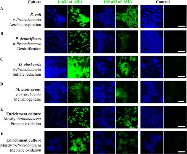
Fig 2.
Uptake and incorporation of AHA is independent of the physiological or phylogenetic background of the target organism. Different pure and enrichment cultures were incubated in the presence or absence of AHA. BONCAT signals (green) were taken at identical exposure times for individual series (i.e. 0.1 and 1 mM AHA plus control). Note that incubation conditions were different for the individual cultures, cells have contrasting levels of background fluorescence, and that different labelling strategies were used. Together, these issues limit the value of a direct comparison of signal intensities between different cultures. DAPI-staining is shown in blue. All scale bars equal 10 μm and apply to each set of images respectively. A–D. BONCAT-labelling of four bacterial and archaeal pure cultures. E–F. BONCAT-labelling of propane- and methane-oxidizing enrichment cultures.

