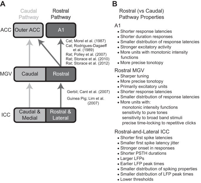
Fig. 12.
Schematic of anatomic projections and summary of physiological differences that indicate the lemniscal pathway is segregated into 2 subprojection pathways. A: the rostral and caudal ascending pathways show spatially segregated anatomic projections from the ICC up to the core auditory cortex regions (ACC). Overlapping projections between the 2 pathways are not shown. A1, primary auditory cortex; MGV, ventral division of the medial geniculate body. B: in contrast to the caudal pathway, the rostral pathway also shows different responses to acoustic stimuli in A1 (Phillips et al. 1995; Polley et al. 2007; Storace et al. 2012; Wallace et al. 2000), rostral MGV (Rodrigues-Dagaeff et al. 1989), and the rostral-lateral ICC (as shown in this study). [Adapted from Lim et al. (2008) with permission.]

