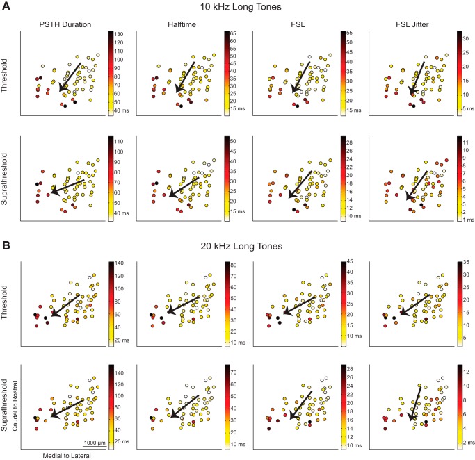
Fig. 4.
Spiking responses to long tones vary from caudomedial to rostrolateral across the lamina. The maps of spiking in response to 10-kHz (A) and 20-kHz long tones (B) show response parameters recorded at each location. Similar to short tone responses in Fig. 3, the responses varied from caudomedial to rostrolateral regions across the lamina.

