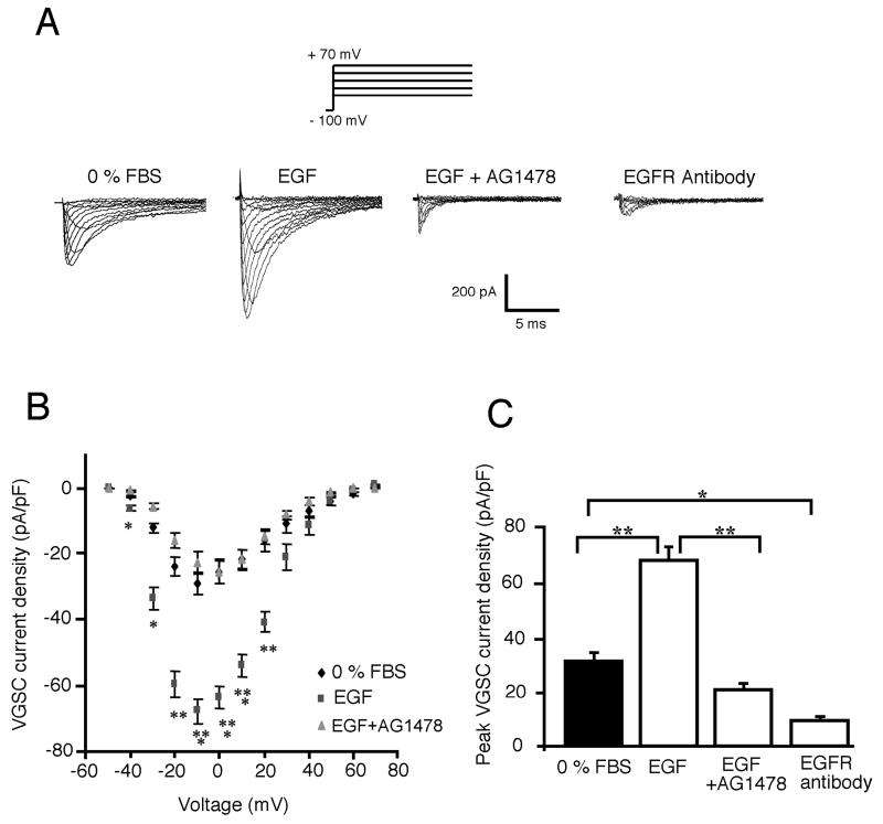
Figure 1. Upregulation of VGSC activity in Mat-LyLu cells by treatment for 24 h with exogenous EGF.
A: Typical VGSC current traces recorded from Mat-LyLu cells in different culture conditions: 0% FBS, EGF (100 ng/ml), EGF + AG1478 (1 μM), and anti-EGFR antibody (1 μg/ml). VGSC currents were recorded by pulsing membrane potentials from −50 to +70 mV in 10 mV increments, from a holding potential of −100 mV. B: Effects of EGF and EGF + AG1478 (as in A) on current–voltage relationship. Peak values of VGSC current density were plotted against membrane potential, showing EGF-stimulated VGSC functional activity. C: Histograms showing mean values of peak VGSC current density recorded in different conditions (as in A). All data points shown are mean and SEM. Significance: *P < 0.05; **P < 0.01; ***P < 0.001.

