
Fig 2.
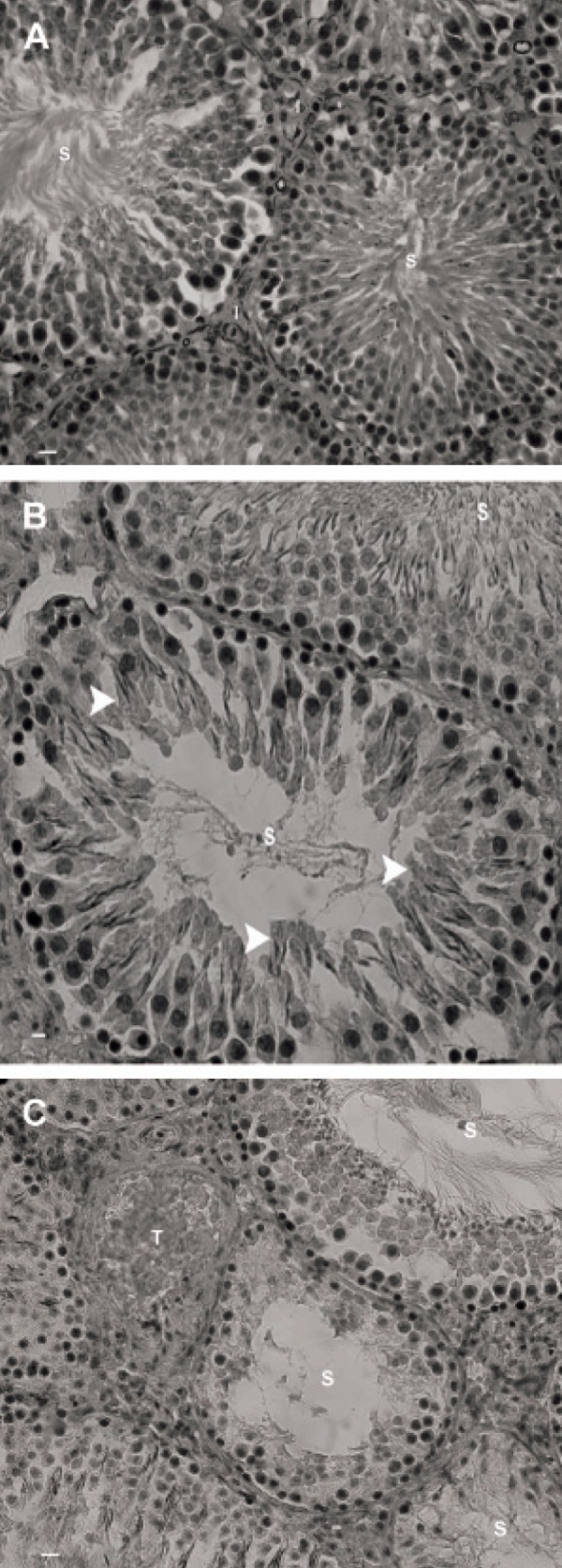
Photomicrograph of rat testis. A. Control-sham group. Note the seminiferous tubules with normal cellular junction (S) and the interstitial connective tissue with no edema (I). B. Right testes of varicocelized rats. Note the seminiferous tubules with negative tubular differentiation index (S). The spermatogenesis regions (arrowheads) are indicative of early maturation from the previous cycle. C. Left testis of varicocelized group. The seminiferous tubules (S) are completely depleted with no detectable spermatic maturation. Vascular thrombosis (T) is also observed in interstitial connective tissue. Iron-Weigert staining, (A × 100; B × 400 and C × 400).