
Fig 4.
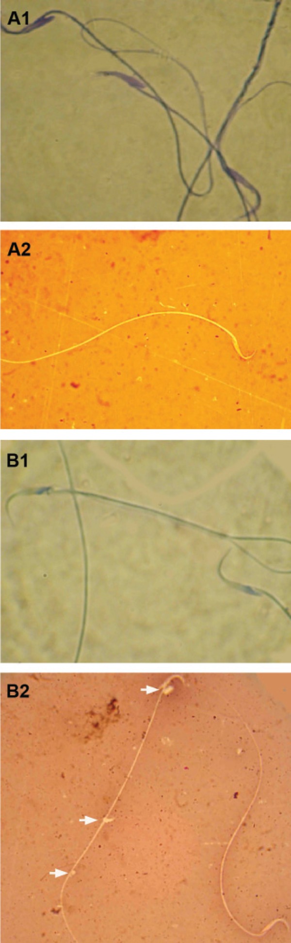
Light microscopic architecture from sperm; A1; Abnormal sperm with dense blue stained mature nucleus, A2; Normal sperm with unstained cytoplasm in head section, B1; Normal sperm with light stained immature nucleus, and B2; Abnormal sperm; note the sperm in the left side of the figure with cytoplasmic droplet (arrows) and the dead sperm with eosin-stained cytoplasm (below, right hand side). Aniline-blue (A-1, B-1) and eosin-negrosin (A-2, B-2) stainings, (× 400).