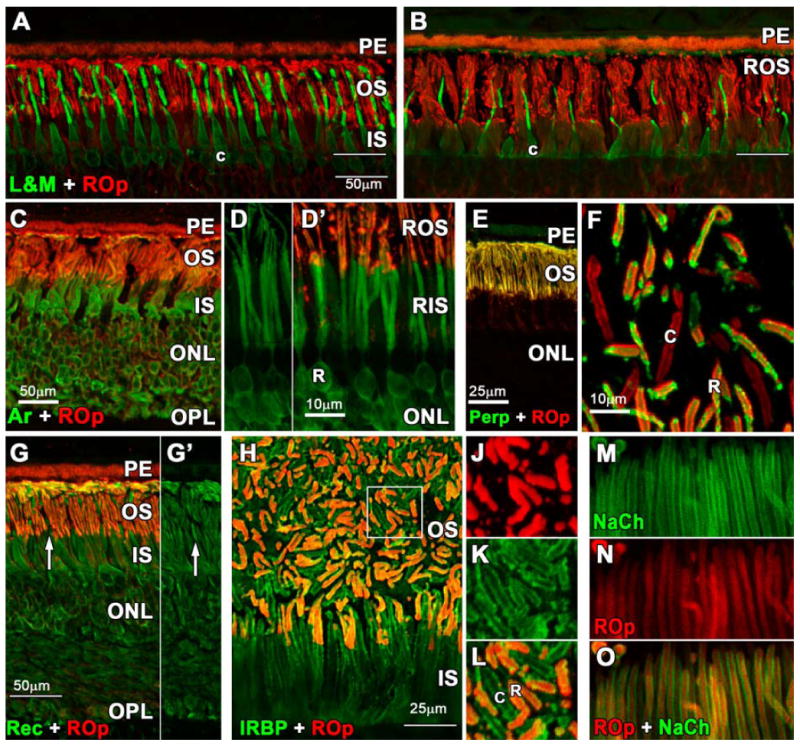
Figure 1.

Photomicrographs of adult human retina immunolabeled with antibodies to rod opsin (ROp, red), L&M cone opsin (L&M; green); rod arrestin, peripherin, recoverin, interphotoreceptor retinoid binding protein (IRBP) and the rod GTP-gated sodium channel (NaCh). A-B. L&M immunoreactive (IR) cones are more numerous and their outer segments (OS) longer in central (A) vs peripheral (B) retina. In central retina the ROp-IR rod OS (red) and L&M cone OS (green) are similar in length. In peripheral retina, cone OS are about half the length of rod OS. White line indicates the external limiting membrane. (Scale bars are 50 μm) C-D′. Arrestin is expressed throughout the rod cytoplasm (C,D) including the synaptic spherules, but arrestin labels the OS relatively lightly compared with ROp (D,D′). E-F. Perpherin is expressed in both rod and cone OS but not the cytoplasm (E). A tangential section shows perpherin-IR cone (F, c) and ROp/Perp-IR rod OS. G. Recoverin is expressed throughout the cytoplasm of both cones (G,G′ white arrow) and rods with relatively light labeling of the OS. H. Double labeling for IRBP (green) and ROp (red) shows that this protein is located on the extracellular OS surface of both rods and cones. The region indicated by the box is shown at higher magnification for J: ROp, K: IRBP, L: ROp/IRBP. M-O. Rod OS have heavy labeling for NaCh in the surface membrane (M) while ROp more evenly labels the OS membrane and discs (N). Scale bars A-C; G = 50μm, D;F;J-K= 10μm, E;H =25μm.
