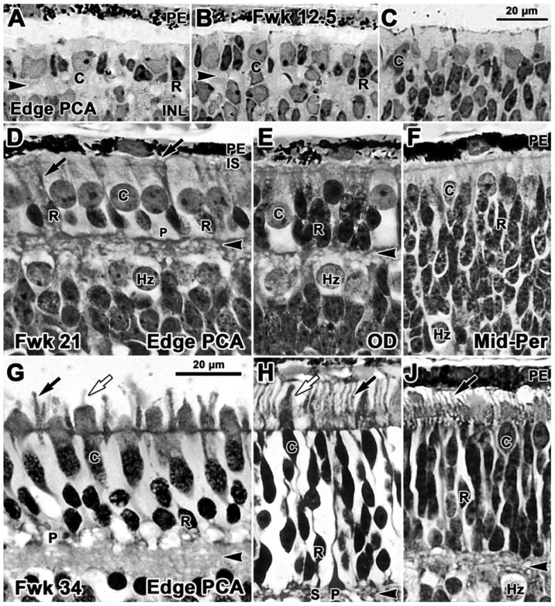
Figure 2.

Photomicrographs from glycol methacrylate sections of human retina stained with azure II & methylene blue. Sections are aligned on the external limiting membrane. A-C Fwk12.5; D-F Fwk21; G-J Fwk34. A. Rods (R) are present on the edge of the pure cone area (PCA), the site of the future fovea. Rods have elongated, densely stained nuclei and a tiny amount of dense cytoplasm in contrast to the pale larger cones (C). B. Rods and cones both are present 400μm further peripheral at the point where the outer plexiform layer (OPL; arrowhead) disappears. C. Only cones can be identified in more peripheral retina. D. Near the developing fovea the smaller dense rod nuclei form a single row of cells adjacent to the OPL. The larger cone nuclei and cell bodies form a packed single layer and cone pedicles (P) line the outer border of the OPL. The inner OPL border is formed by a band of pale horizontal cells (Hz). Thin dense inner segments (IS; arrow) are present on rods. OS cannot be easily identified, although antibody labeling (see Fig.6) shows that they are present on both cones and rods. E. Temporal to the optic disk (OD) cones are sparse while rods are stacked 3-4 cells deep adjacent to the thin OPL. F. In midperipheral retina the OPL is not yet present. A single row of cones lines the outermost retina, while rods are stacked 10-12 cells deep and merge with pale Hz. G. Near the fovea, rods are 2 cells deep and have well developed IS and OS (black arrow). Cones have thick IS and short OS (white arrow). H. At the OD rods greatly outnumber cones. Compared to G, both rods and cones are more mature, have longer IS and OS and distinct synaptic spherules (S) and pedicles (P). J. A thin OPL is present in the midperiphery and rods are stacked 9-12 cells deep under the cones. Both rods and cones have short IS and OS (arrow). Scale bar in C for A-C; in G for D-J.
