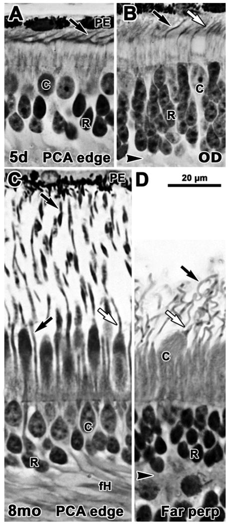
Figure 3.

Photomicrographs from glycol methacrylate sections of human postnatal retina. Sections are aligned on the external limiting membrane. A-B 5 days; C-D 8 months. A. Near the PCA rods are 2-3 deep with distinct OS present on both rods (arrow) and cones. B. Near the OD rods are 5-6 cells deep and both rods (black arrow) and cones (white arrow) have significantly longer IS and OS than those on the edge of the fovea. C. Rod (black arrow) and cone (white arrow) OS on the PCA edge are much longer than in the newborn. Photoreceptors in and around the fovea have also elaborated long basal axons (fH), increasing the thickness of the OPL. D. Very long, thin rod OS and shorter, thicker cone OS are present well into the periphery. Scale bar in D for A-D.
