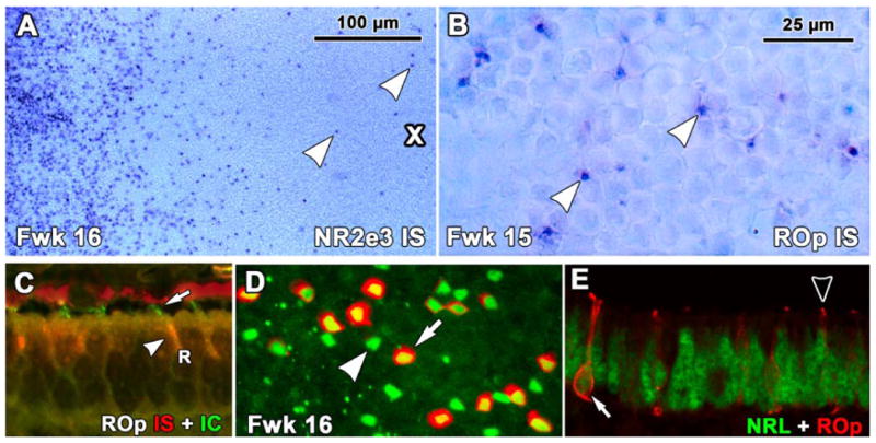
Figure 5.

Expression of rod opsin (ROp) mRNA as detected by insitu hybridization (IS) in human fetal retina. A. A Fwk16 wholemount hybridized to detect NR2E3 mRNA around the PCA (cross). The PCA is surrounded by a thick band of NR2E3 expressing rods with scattered NR2E3 expressing rods within the PCA (arrowheads). B. ROp mRNA is first expressed in scattered rods on the edge of the PCA (arrowheads) C. Fwk16 section processed first for ROp mRNA (red) and then for ROp protein (green). Rods have red IS (arrowheads) and short green OS (arrow). D: A Fwk16 wholemount at the edge of ROp expression double labeled for NRL (green) and ROp protein (red). Note the large number of single labeled NRL-IR nuclei (arrowhead) with NRL/ROp-IR rods scattered in between (arrow). E, A Fwk16 section at the front of ROp expression. The arrow indicates a rod with a NRL-IR nucleus (green) which is outlined by ROp-IR membrane (arrow, red). Most other adjacent rods have either NRL-IR nuclei only or a tiny ROp-IR OS (arrowhead). Scale bar in B for B-E.
