
Figure 6.
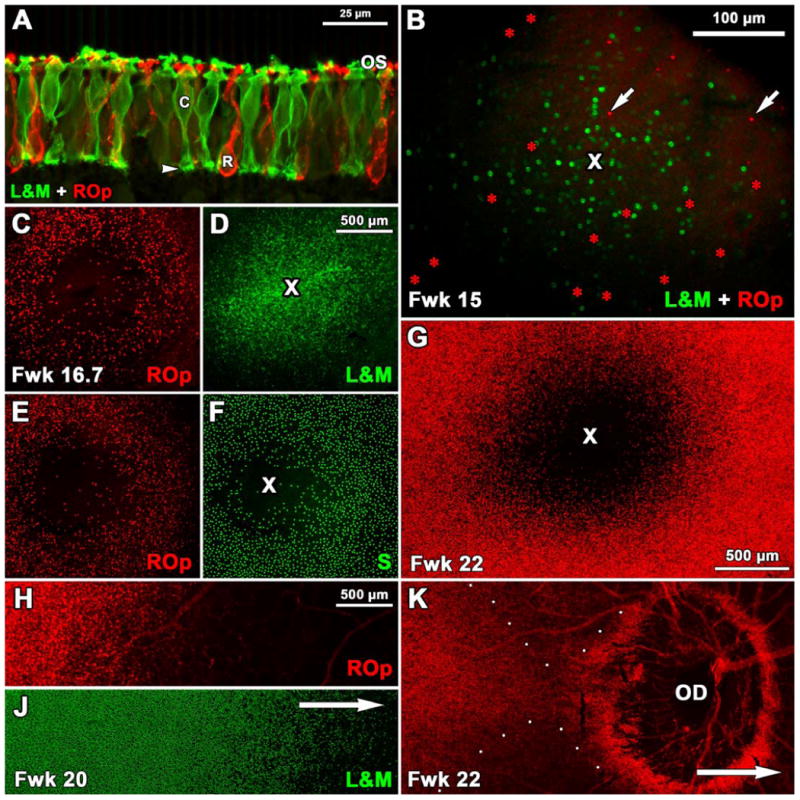
Expression of ROp, L&M and S opsin in fetal human retina. A: A section through the PCA edge at Fwk 18 shows many ROp-IR rods (red) and L&M-IR cones (green). The entire photoreceptor cell membrane including the synaptic pedicle of cones (arrowhead) is labeled. Short OS are present on both rods and cones. B. Fwk15 wholemount showing 262 L&M-IR (green) cones and 37 ROp-IR (red, arrows) rods present in or around the PCA (cross). C-F: Two retinal wholemounts from the same Fwk16.7 fetus. ROp labeling (D,F, red) shows a 680μm wide PCA which not entirely “rod-free”. The future foveal center is packed with L&M-IR cones (E, cross) and is surrounded by a dense ring of S-IR cones which are sparse in the PCA center (F, cross). G: By Fwk 22 the PCA is almost completely rod free, and is surrounded by densely packed ROp-IR rods. H-J. Double labeled Fwk 20 wholemount showing the relative position for the fronts of ROp (J) and L&M (K) expression. Arrow indicates direction of the optic disc (OD). K: Wholemount showing precocious ROp expression (red). Although the main expression front (to the left) has not reached the OD, many ROp-IR rods surround it. ROp-IR rods are absent peripheral to the OD (direction of arrow). Scale bar in D for C-F; in H for H-K.