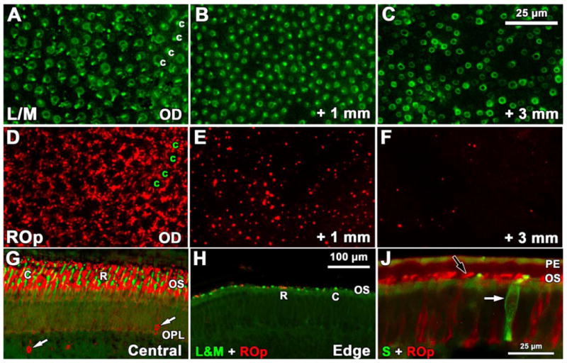
Figure 7.

Development of rod and cone opsin labeling in late fetal and neonatal human retina. A-F: Fwk 25 wholemount showing the pattern and relative number of L&M-IR cones (A-C) and ROp-IR rods (D-F) near the optic disc (A,D), 1mm (B,E) and 3mm (C,F) peripheral to the optic disc. A,D. Near the optic disc L&M-IR cones (A, c) are encircled by a dense rim of ROp-IR rods (D) in a mature pattern, suggesting that all photoreceptors have expressed opsin. B, E. At 1mm peripheral to the optic disc the L/M-IR cone pattern (B) has a few regions lacking cones, but rod density has dropped markedly (E). C,F. At 3mm there are fewer L/M-IR cones (C) although they can be identified at least several mm more peripherally. Rods (F) are very sparse at 3mm G-H. A comparison of ROp and L/M labeling in central retina (G) and near the retinal edge (H) at P1day. The neonatal central retina has long cone (c; green) and rod (r; red) OS while both are sparse and very short at the retinal edge. The central retina has many ROp-IR ectopic cells in inner retina (arrows). J: A comparison of ROp-IR (black arrow) rods and S opsin-IR cones (white arrow) in the periphery of the 5day retina. Scale bar in C for A-F; bar in H for G-H.
