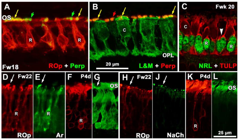
Figure 8.

Expression of phototransduction and OS structural proteins. A-B. Sections near the Fwk18 PCA. A shows rods double-labeled for peripherin (perp; green) and ROp (red) while B shows cones double labeled for L&M (green) and peripherin (perp; red). Both rod and cone OS are double labeled (yellow arrows) while peripherin-IR cone OS (A, green arrows) and rod OS (B, red arrows) are intermixed. Perpipherin label in cell bodies is minimal. Note that rods lack any obvious synaptic spherule but cones have a well developed pedicle in the OPL. C: Section of Fwk20 retina near the PCA edge showing the appearance of TULP (red) in rods which have an NRL-IR nucleus (green). A thin rim of TULP-IR cytoplasm (arrowhead) marks the rods. Cone cytoplasm is also TULP-IR but OS do not label. D-E: Fwk22 rods OS double label for ROp (D) and rod arrestin (E). Both the cell membrane and short OS (arrow) double label. F-G. Postnatal 4day rods have mainly OS label for ROp (F) with heavy cytoplasmic and lighter OS labeling for arrestin (G). The arrow indicates an arrestin-IR S cone OS. H-J. Most of the short Fwk22 rod OS (arrows) are double labeled for ROp and NaCh. K-L. Rod OS labeling has increased at 4days for ROp (K) and NaCh (L) with little cytoplasmic labeling for either protein. Magnification bar in B for A-B; bar in L for D-L.
