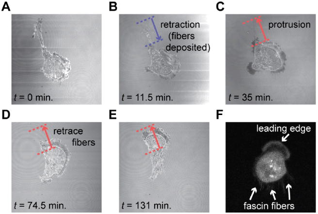
Fig. 6.

Confocal microscopy in reflection mode of a MDA-MB-231 cell leaving a bundle of fibers on the glass substrate (coated with laminin) after a major tail retraction. (A) The cell retracts its tail along the blue arrow. (B) After retraction of the tail, a “fiber trail” consisting of multiple thin retraction fibers remains on the substrate. (C) Shortly thereafter, the cell retraces its path along the fiber trial. (D–E) The deposited fibers are removed after the cell retraces its previous path and uptakes the fibers. (F) MDA-MB-231 cell transfected with fascin-GFP (confocal microscopy image) showing that the retraction fibers are rich in fascin. See also ESI† Movie M6.
