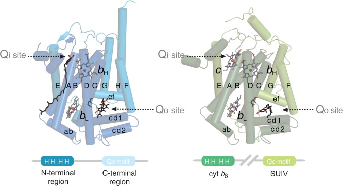
Fig. 2.—

Structural composition of cyt b. The subunit can be divided in N- and C-terminal regions. Cyt b is encoded as single protein in mitochondria and most bacteria (left, pdb 2ibz). In cyt b6f complexes, these two regions are encoded as two separate polypeptides (right, pdb 1q90) and are denoted cyt b6 and SUIV. Transmembrane and relevant surface helices are labeled with upper and lower case letters, respectively. The Qi site of cyt bc1 complexes is occupied by the natural substrate ubiquinone-6. The Qo site of both cyt bc1 and cyt b6f complexes are, respectively, indicated by their inhibitors, stigmatellin and tridecylstigmatellin, in hydrogen bonding distance to the Glu272 equivalent residue of the Qo motif. The structural composition of cyt b is shown schematically in the lower panel. The four “H” denote the four strictly conserved histidine axial ligands of heme bH and heme bL, and the Qo motif is labeled on the C-terminal region and SUIV. Cyt b6f complexes possess a heme ci at the Qi site, which is absent in cyt bc1 complexes.
