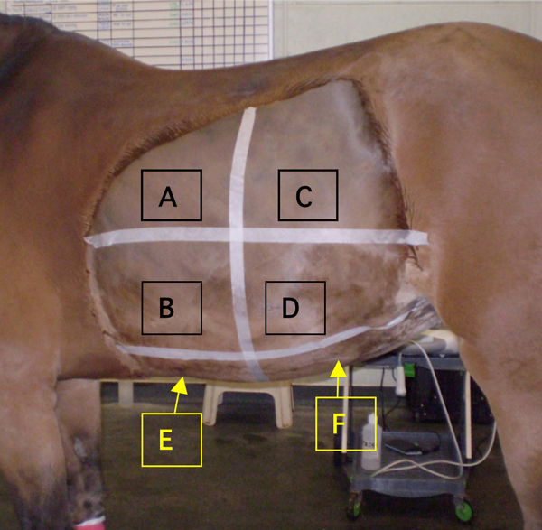
Figure 1.

Photograph of the left abdomen illustrating the six regions for ultrasonographic examination. Sites A, B, C and D were repeated on the right abdomen. A: Left craniodorsal flank, B: Left cranioventral flank, C: Left caudodorsal flank, D: Left caudoventral flank , E: Cranioventral, F: Caudoventral
