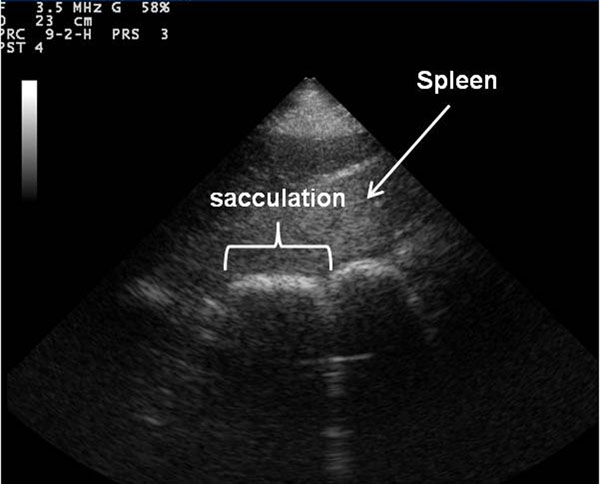
Figure 2.

Ultrasonographic image of sacculated large intestine. Frontal plane ultrasound image of sacculated large intestine with cranial to the left of the image obtained in the left ventral region of the abdomen using a detailed transcutaneous ultrasound technique.
