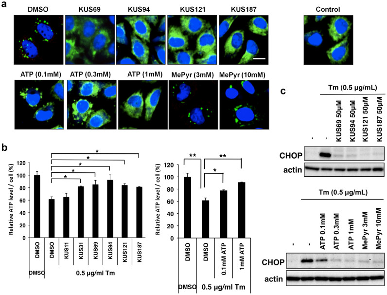
Figure 4. KUSs and ATP each prevented a decrease of ATP levels and ameliorated ER stress in tunicamycin-treated cells.
(a) Immunocytochemical analyses of HeLa cells by an anti-laminin γ1 antibody. HeLa cells were treated with 0.5 μg/ml of tunicamycin (Tm) for 5 hours in the presence of KUSs (50 μM), ATP (0.1, 0.3, and 1 mM), methylpyruvate (MePyr) (3 and 10 mM), or vehicle alone (DMSO). Then, cells were fixed and subjected to immunocytochemical analyses. Normally growing HeLa cells were also analyzed (Control). Scale bar, 10 μm. (b) Measurements of the relative amounts of ATP per cell. HeLa cells were treated with tunicamycin (Tm, 0.5 μg/ml) for 24 hours in the presence of KUSs (50 μM) or ATP (0.1 and 1 mM), or vehicle alone (DMSO), and were harvested. Then, ATP amounts from 1.5 × 10⁵ cells were measured27. * P < 0.05, ** P < 0.01. Error bars indicate SD. (c) Western blot analyses on CHOP. HeLa cells were treated with 0.5 μg/ml of tunicamycin for 5 hours in the presence of KUSs (50 μM), ATP (0.1, 0.3, and 1 mM), methylpyruvate (MePyr) (3 and 10 mM), or vehicle alone (-). Then, cells were harvested and subjected to western blot analyses. Actin served as a loading control. Complete scans of the different blots are presented in Supplementary Fig. 9.

