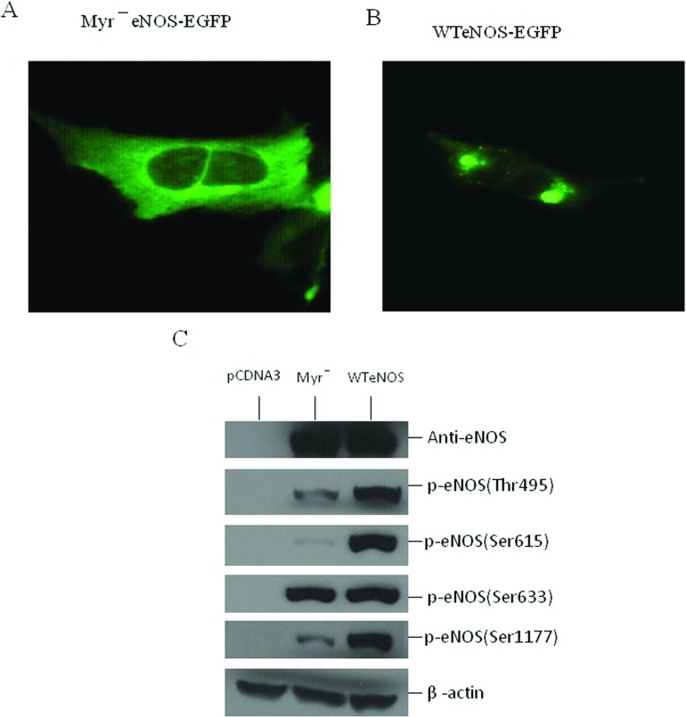
Figure 2. Relationship between the subcellular localization and phosphorylation state of eNOS.
Stable HEK-293 cell lines with (A) myr−eNOS-EGFP or (B) WTeNOS–EGFP were visualized in live cells by fluorescence microscopy. (C) cell lysates from HEK-293 cells transfected with empty vector (pCDNA3), Myr−eNOS or WTeNOS were immunoblotted for changes in phosphorylation state of Thr495, Ser615, Ser633 and Ser1177. The expression of eNOS and β-actin was used as internal loading control. Western blots shown are the representatives of at least three independent studies.

