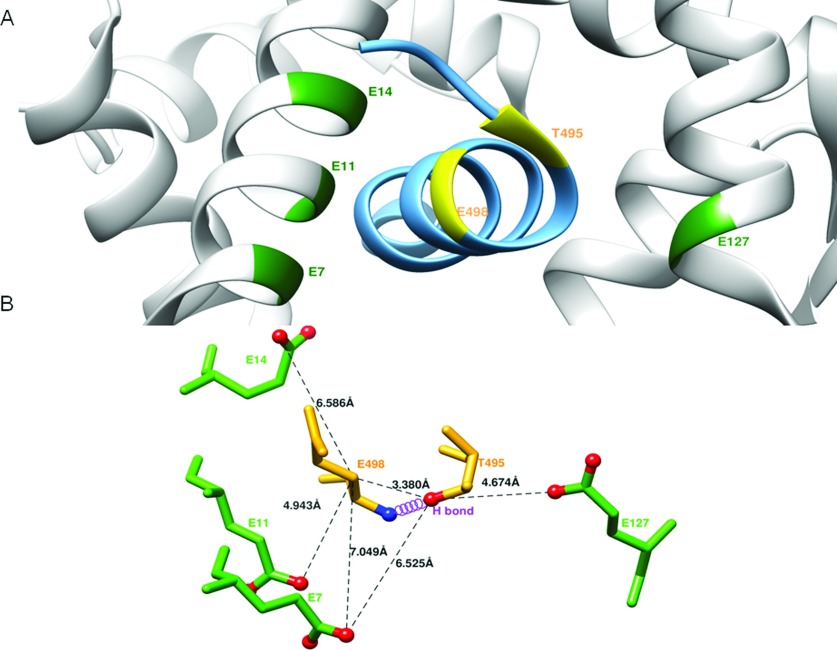
Figure 7. T495 phosphorylation and eNOS function.
(A) Structure of CaM and its complex with eNOS CBD (PDB accession code 1NIW). The backbone ribbons of the CBD (sky blue) and of CaM (grey) are shown. The T495 and E498 in eNOS CBD are labelled in yellow, and the neighbouring glutamate residues located on CaM in the vicinity of Thr495 and Glu498 including E7, E11, E14 and E127 are labelled in green. (B) Close-up view showing residues surrounding the T495 phosphorylation site. The residues as ball-and-stick representation are coloured by atom type (nitrogen, blue; oxygen, red). The hydrogen bond between Thr495OG1 and Glu498 amide N is indicated by the violet coil. The selected distances between residues are measured from the structure (1NIW) and indicated in dash lines: Thr(495)Oγ-Glu(498)Cβ, 3.38 Å; Thr(495)Oγ-Glu(7)Oε, 6.53 Å; Thr(495)Oγ-Glu(127)Oε, 4.67 Å; Glu(498)Cβ-Glu(7)Oε,7.05Å; Glu(498)Cβ-Glu(11)Oε,4.94Å; Glu(498)Cβ-Glu(14) Oε, 6.59Å.

