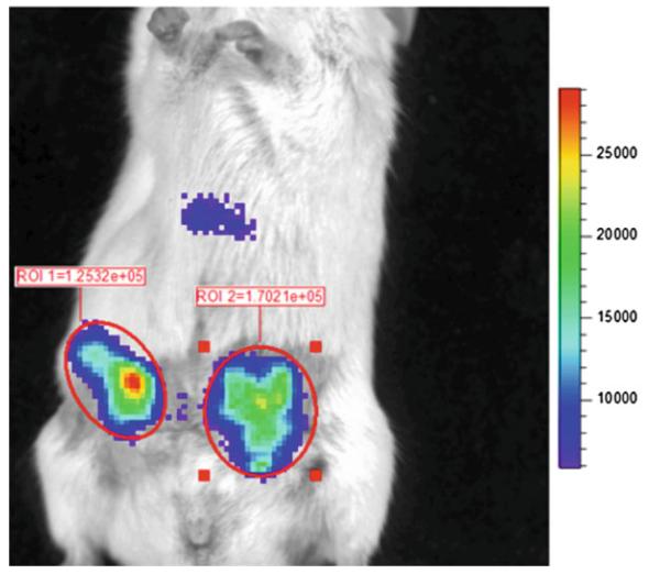
Fig. 3.
Imaging ligand–receptor binding in living mice. Mice were implanted with equal numbers of 231-CXCL12-CGLuc and 231-NGLuc-CXCR4 cells as ortho-topic mammary tumor xenografts in NSG mice. Imaging began 20 s after tail vein injection of coelenterazine, using 2 min acquisition and large binning. Circles and values show photon fl ux measurements for ROIs around each tumor. Scale bar shows range of values depicted by pseudocolor display

