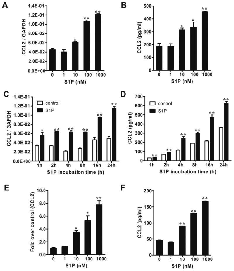
Figure 1.

S1P induced CCL2 expression in NB cells. (A and B) SK-N-AS cells were serum starved for 24 h and treated with different concentrations of S1P for 4 h followed by quantitative real-time PCR (A) and CCL2 ELISA (B). (C and D) Serum-starved SK-N-AS cells were treated with 1μM of S1P for 1-24 h followed by quantitative real-time PCR (C) and CCL2 ELISA (D). (E and F) SK-N-BE(2) cells were serum starved for 24 h and treated with S1P for 2 h (E) or 4 h (F) followed by quantitative real-time PCR (E) and CCL2 ELISA (F). *, P < 0.05, **, P < 0.01 versus without S1P treatment.
