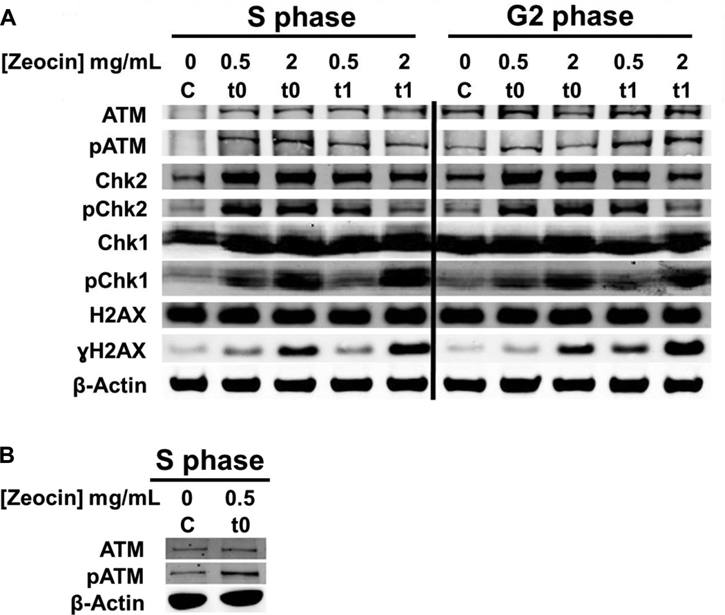
Fig. 2. Activation of the DNA damage response pathways in S and G2.
(A) Cells were synchronized by a double thymidine block and released for 3 and 7 hours (S and G2 cells, respectively). These cells were treated with Zeocin™ for 1 h and harvested at t0 and one hour post treatment (t1). Whole cell lysates were analyzed by western blot with indicated antibodies (50 µg whole cell lysate per lane). (B) Repeat of ATM and pATM western blots in S phase for control and t0 to show that the low recognition of ATM in the control sample of panel A are putatively due to reduced transfer of high molecular weight proteins in that part of the gel.

